Home » Without Label » Arteries In Neck - Circulation Of The Internal Carotid Artery Modified From Netter Fh Download Scientific Diagram - There are two large arteries in the neck, one on each side.
Arteries In Neck - Circulation Of The Internal Carotid Artery Modified From Netter Fh Download Scientific Diagram - There are two large arteries in the neck, one on each side.
Arteries In Neck - Circulation Of The Internal Carotid Artery Modified From Netter Fh Download Scientific Diagram - There are two large arteries in the neck, one on each side.. With a cervical artery dissection, the neck pain is unusual, persistent, and often accompanied by a severe headache, says dr. The carotid arteries are major blood vessels in the neck that supply blood to the brain, neck, and face. The vertebral arteries ascend through the neck inside the transverse foramina of the cervical vertebrae, all the way to the brain. Without this blood flow, your brain cells would. Blood flow in this artery can become partly or totally blocked by fatty material called plaque.
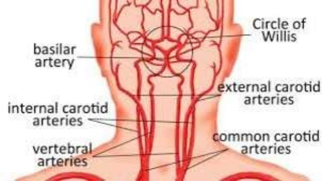
However, neck arteries can work just as fine, even though they are partially blocked. The vertebral arteries terminate by anastomosing together as the basilar artery. Carotid arteries are located in the anterior of the neck, on either side. A stroke can occur when your carotid arteries become blocked.read more. However, pain from carotidynia typically only occurs on one side.
Carotid Arteries from www.thoughtco.com They are the carotid arteries, and they carry blood to the brain. Your healthcare provider uses a device called a transducer to make pictures of the arteries. The vertebral arteries terminate by anastomosing together as the basilar artery. Blood is carried to the brain through blood vessels called arteries. The arteries in neck that supply blood to the brain are called carotid arteries. The carotid arteries can be felt on each side of the lower neck, immediately below the angle of the jaw. Your doctor may then test your physical and mental capabilities such as strength, memory and speech. Carotid artery disease occurs when fatty deposits (plaques) clog the blood vessels that deliver blood to your brain and head (carotid arteries).
The arteries in the chest, neck and brain are the most frequent arteries found to be abnormal in phace syndrome.
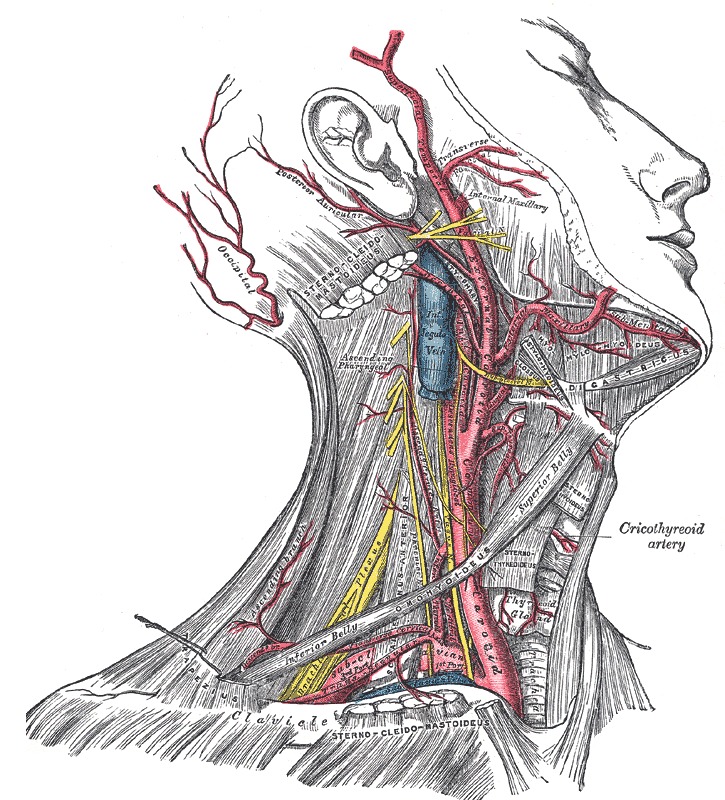
The internal carotid artery is a major contributor to the circulus arteriosus or circle of willis which supplies the greater part of the brain. Our technicians use an ultrasound machine to scan the carotid artery and to check for any plaque buildup in this critical area. Doctors can test for a narrowed carotid artery, but it's usually not a good idea. A condition which arises spontaneously or as the result of trauma, where the walls of the artery are split, leading to internal bleeding and disruption of blood flow. At the root of the neck the right internal jugular vein is placed at a little distance from the common carotid artery, and crosses the first part of the subclavian artery, while the left internal jugular vein usually overlaps the common carotid artery. At the level of the superior margin of the thyroid cartilage (c4), the carotid arteries split into the external and internal carotid arteries. Though more often occurring with carotid arteries (the other major ones supplying the brain through the neck), vertebral arteries can be impacted. These blood vessels can have abnormal shapes, sizes or paths through the neck and head. They are called the carotid arteries, to be more precise the external and the internal carotid artery. Veins and arteries of the neck 9 photos of the veins and arteries of the neck activate javascript arteries in the neck diagram, common carotid artery branches, external carotid artery function, how many carotid arteries, left common carotid artery function, the left common carotid artery supplies blood to the. The plaque buildup is made of fat, cholesterol, cellular waste, calcium, proteins and inflammatory cells. The exam generally includes listening for a swooshing sound (bruit) over the carotid artery in your neck, a sound that's characteristic of a narrowed artery. Your doctor may then test your physical and mental capabilities such as strength, memory and speech.
Related posts of arteries in the neck picture veins and arteries of the neck. At the root of the neck the right internal jugular vein is placed at a little distance from the common carotid artery, and crosses the first part of the subclavian artery, while the left internal jugular vein usually overlaps the common carotid artery. The arteries in the chest, neck and brain are the most frequent arteries found to be abnormal in phace syndrome. Doctors can test for a narrowed carotid artery, but it's usually not a good idea. They are the carotid arteries, and they carry blood to the brain.
Arteries Of Neck from image.slidesharecdn.com The neck pain from a carotid artery tear often spreads along the side of the neck and up toward the outer corner of the eye. Your doctor may then test your physical and mental capabilities such as strength, memory and speech. A condition which arises spontaneously or as the result of trauma, where the walls of the artery are split, leading to internal bleeding and disruption of blood flow. Just like other arteries in the body, neck arteries are also susceptible to blockages. Blood is carried to the brain through blood vessels called arteries. Blood flow in this artery can become partly or totally blocked by fatty material called plaque. The left and right common carotid arteries ascend up the neck, lateral to the trachea and the oesophagus. Without this blood flow, your brain cells would.
These blood vessels can have abnormal shapes, sizes or paths through the neck and head.
Carotid artery disease occurs when fatty deposits (plaques) clog the blood vessels that deliver blood to your brain and head (carotid arteries). The exam generally includes listening for a swooshing sound (bruit) over the carotid artery in your neck, a sound that's characteristic of a narrowed artery. The artery walls are made up of three layers of different types of tissue, each with a specific function. In the neck, the carotid sheath (fibrous connective tissue) covers the common carotid artery, vagus nerve, and internal jugular vein. This can reduce the blood supply to your brain and cause a stroke. Carotid arteries are located in the anterior of the neck, on either side. What are the carotid arteries? They do not give off any branches in the neck. What is a carotid artery duplex scan? Our technicians use an ultrasound machine to scan the carotid artery and to check for any plaque buildup in this critical area. If one of them is narrowed or blocked, it can lead to a stroke. The pain may be sudden and severe—people often describe it as a throbbing pain. The carotid arteries are two large blood vessels that supply oxygenated blood to the large, front part of the brain.
Veins and arteries of the neck 9 photos of the veins and arteries of the neck activate javascript arteries in the neck diagram, common carotid artery branches, external carotid artery function, how many carotid arteries, left common carotid artery function, the left common carotid artery supplies blood to the. The carotid artery brings needed blood to your brain and face. This can reduce the blood supply to your brain and cause a stroke. Carotid artery disease is when fat accumulates and blocks the blood flow of your neck arteries (carotid arteries).your carotid arteries supply your brain with blood rich in oxygen. A vertebral artery tear may feel like something sharp is stuck in the base of your skull.
Figure Arteries Of The Head And Statpearls Ncbi Bookshelf from www.ncbi.nlm.nih.gov Carotid artery disease is when fat accumulates and blocks the blood flow of your neck arteries (carotid arteries).your carotid arteries supply your brain with blood rich in oxygen. The plaque buildup is made of fat, cholesterol, cellular waste, calcium, proteins and inflammatory cells. You have one of these arteries on each side of your neck. Without this blood flow, your brain cells would. The vertebral arteries terminate by anastomosing together as the basilar artery. Stroke deprives your brain of oxygen. What are the arteries of the chest, neck and brain? There are two large arteries in the neck, one on each side.
Blood is carried to the brain through blood vessels called arteries.
In the neck, the carotid sheath (fibrous connective tissue) covers the common carotid artery, vagus nerve, and internal jugular vein. The vertebral arteries terminate by anastomosing together as the basilar artery. This is where thinking, speech, personality, and. The exam generally includes listening for a swooshing sound (bruit) over the carotid artery in your neck, a sound that's characteristic of a narrowed artery. With a cervical artery dissection, the neck pain is unusual, persistent, and often accompanied by a severe headache, says dr. The vertebral arteries ascend through the neck inside the transverse foramina of the cervical vertebrae, all the way to the brain. Your carotid arteries are the major blood vessels that deliver blood to your brain. This artery also brings blood to your brain. The test may also look at the vertebrobasilar artery. What are the arteries of the chest, neck and brain? Carotid artery disease occurs when fatty deposits (plaques) clog the blood vessels that deliver blood to your brain and head (carotid arteries). The carotid arteries can be felt on each side of the lower neck, immediately below the angle of the jaw. There are two carotid arteries, one on the right and one on the left.
/carotid-neck-3ab3e1120b654a24af1a35ddf0e267f6.jpg)